Case 11 — Thoracic Radiographs
DV

Right Lateral
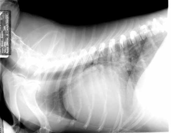
One of the primary things one would like to evaluate on this dog is its lung fields for the presence of pulmonary edema. Unfortunately, this dog’s diaphragm is displaced forward by its ascites, making it impossible to tell if there are infiltrates in the caudodorsal lung fields. If there is pulmonary edema, it would most likely be mild to moderate. At this stage, pulmonary edema is usually centrally located on the DV radiograph and consequently mild to moderate edema usually cannot be identified in this view.