Skip to main content
SACM
Home
Cases
ECG Atlas
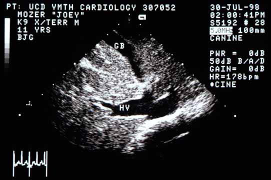
Case 11 — Abdominal Ultrasound
Abdominal ultrasound of the liver. The proximal hepatic vein (HV) and caudal vena cava are markedly distended, typical of congestive right heart failure. GB — gall bladder.
← Doppler
Next: Diagnosis →
↑