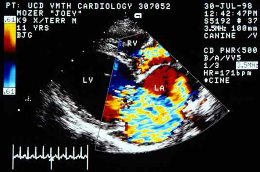
Case 11 — Color Flow Doppler Echocardiography

Color flow Doppler image in systole showing blood flowing from the left ventricle (LV) into the left atrium (LA). The jet is large, extending into the distended pulmonary vein. There is also a color jet emanating from the right ventricle (RV) into the right atrium, indicating tricuspid regurgitation.
Systolic color flow Doppler. A large MR jet extends from the LV into the LA and the distended pulmonary vein. A TR jet also emanates from the RV into the RA.
Color flow Doppler image showing blood flowing from the left atrium (LA) through the defect in the atrial septum into the right atrium (RA) during systole. PV — pulmonary vein; AO — aorta; PA — pulmonary artery.
Systolic color flow Doppler showing blood flowing from the LA through the atrial septal defect into the RA. PV; AO; PA.