Case 11 — M-Mode Echocardiogram
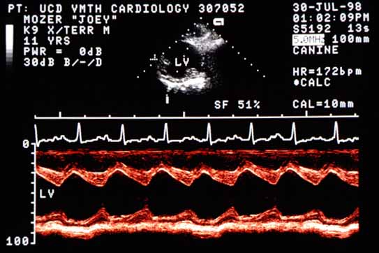
M-mode echocardiogram showing hyperdynamic left ventricular function. End-diastolic diameter is 4.7 cm (normal ~3 cm for this size dog). End-systolic diameter is 2.3 cm (indistinguishable from normal). Shortening fraction is increased at 51%.
M-mode echocardiogram. EDD = 4.7 cm (normal ~3 cm); ESD = 2.3 cm (normal range). Shortening fraction = 51% (increased). Walls move 2.4 cm closer together (normal ~1 cm).

This M-mode demonstrates hyperdynamic left ventricular function. The walls move more than normal, with the interventricular septum and left ventricular free wall moving 2.4 cm closer together from end-diastole to end-systole (normal would be about 1 cm).


Quiz

1. The fact that this dog’s end-systolic diameter is normal tells you what?

2. The increased shortening fraction with a larger than normal diastolic diameter does not tell you what?