Case 6 — Cardiac Catheterization and Angiography

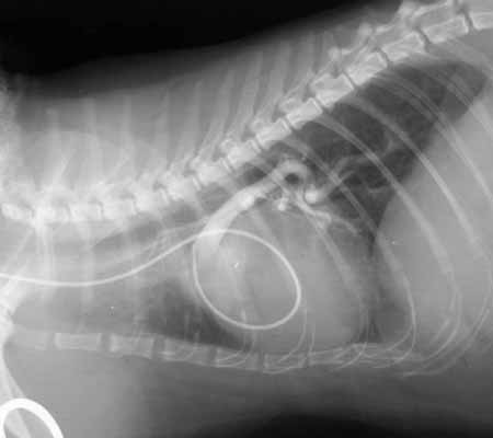
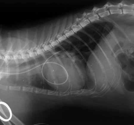
At cardiac catheterization, a 4 French balloon wedge catheter was advanced from the jugular vein into the right heart. Initially, the catheter was advanced into one position (left image below). When it was withdrawn and advanced again, it went into a totally different position that was more dorsal than the first. It appeared that the catheter initially advanced into the pulmonary artery and the second time advanced into the aorta.
Fluoroscopic image showing the balloon wedge catheter advanced into the first position, consistent with the pulmonary artery.
First catheter position (pulmonary artery).
Fluoroscopic image showing the catheter advanced into a second, more dorsal position consistent with the aorta after being withdrawn and readvanced.
Second catheter position (aorta) after withdrawal and readvancement.
Angiogram obtained after injecting nonionic contrast agent from the catheter positioned in the pulmonary artery. The image shows an enlarged and very tortuous caudal lobar pulmonary artery branch.
The catheter was partially withdrawn toward the pulmonary artery and nonionic contrast was injected. The angiogram shows an enlarged and very tortuous caudal lobar pulmonary artery branch.
Angiogram after repositioning the catheter into the structure (S) between the aorta and the main pulmonary artery. Contrast fills the structure, the main pulmonary artery back to the pulmonic valve and its caudal lobar branches, and the aorta from the aortic root to the diaphragm.
Angiogram with catheter in the structure (S). Contrast fills the structure, the main pulmonary artery (MPA) and its branches, and the aorta (Ao) from root to diaphragm.
Labeled version of the same angiogram, identifying the structure (S) between the main pulmonary artery (MPA) and the aorta (Ao).
Labeled version of the same angiogram.

Quiz

1. What is the structure?