Case 6 — Intracardiac Pressures

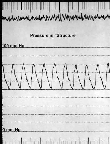
The catheter was passed into the aorta from the pulmonary artery and pressures were recorded as the catheter was withdrawn from the aorta, through the unidentified structure, into the pulmonary artery, and then into the right ventricle. As can be seen below, systolic pressure was equal in all of these structures. Systolic and diastolic pressures were equal in the aorta, in the pulmonary artery, and in the “structure.” The right ventricular systolic pressure was the same as in the aorta, the pulmonary artery, and the structure. The right ventricular diastolic pressure was mildly increased to about 7 mmHg (normal is 5 mmHg or less).
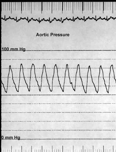
Pressure tracing recorded from the aorta showing systolic and diastolic waveforms. Systolic pressure is elevated and matches pressures recorded from other structures.
Aortic pressure tracing.
Pressure tracing recorded from within the ductus (structure S), showing systolic and diastolic pressures equal to those in the aorta and pulmonary artery.
Ductus pressure tracing.
Pressure tracing recorded from the pulmonary artery as the catheter was withdrawn from the ductus into the PA. Systolic pressure remains equal to aortic pressure, confirming pulmonary hypertension.
Pulmonary artery pressure tracing.
Pressure tracing recorded from the right ventricle. Systolic pressure equals aortic pressure. Diastolic pressure is mildly elevated to approximately 7 mmHg (normal is 5 mmHg or less).
Right ventricular pressure tracing. Diastolic pressure mildly elevated (~7 mmHg).