Case 6 — Echocardiography

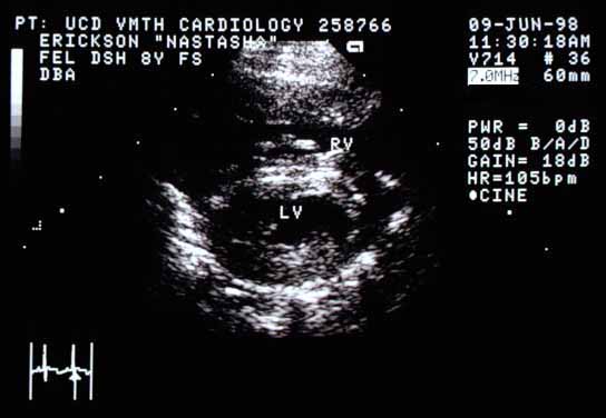
Right parasternal long-axis echocardiographic view of Nastasia's heart. The left ventricular cavity (LV) is smaller than normal. The right ventricular cavity (RV) is visible.
Right parasternal long-axis view. The left ventricular cavity (LV) is smaller than normal. RV — right ventricular cavity.
Right parasternal cross-sectional echocardiographic view (90-degree rotation from the long-axis view). The left ventricular chamber (LV) is again small. The right ventricular free wall (RV) appears markedly thickened.
Right parasternal cross-sectional view (90° rotation from the image above). The left ventricular chamber (LV) is again small. RV — right ventricular chamber.

Quiz

1. What cardiac structure is markedly thickened?

2. What would be the most common pathophysiologic reason for this thickening?