Case 6 — Echocardiograms and Doppler Studies
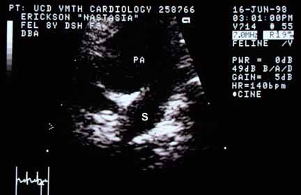
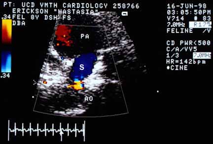
A contrast echocardiogram was performed by injecting agitated saline into a peripheral vein and watching the microbubbles traverse the heart. The microbubbles appeared in the right heart but none were seen in the left heart or aorta. Subsequently, the ultrasound transducer was placed over the caudal abdomen and the aorta identified above the bladder. Agitated saline was again injected. No microbubbles were identified in the caudal aorta, but the image was not perfect and so some doubt existed as to the validity of this test. Further echocardiographic studies were then undertaken.