Case 6 — Radiographs

DV

Dorsoventral (DV) thoracic radiograph showing an abnormally shaped cardiac silhouette with right-sided enlargement including an enlarged main pulmonary artery and aorta. Lung fields are clear with no evidence of pulmonary edema.
Dorsoventral (DV) thoracic radiograph.

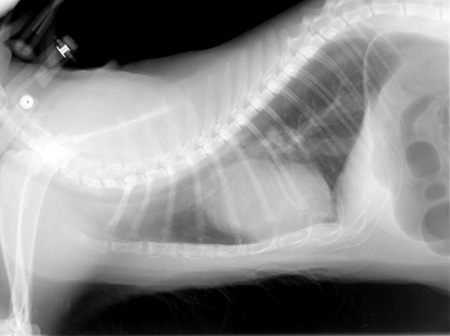
Right Lateral

Right lateral thoracic radiograph showing an enlarged cardiac silhouette with right-sided enlargement and an enlarged proximal descending aorta. The caudal vena cava is also enlarged. Lung fields are clear.
Right lateral thoracic radiograph.

Quiz

1. The heart is abnormally shaped. What enlargement(s) is/are causing this?

2. Is there evidence of pulmonary edema?

3. What great vessel is enlarged besides the aorta?