Case 7 — Case Discussion
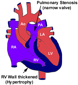
Diagram of pulmonic stenosis.
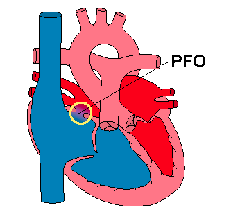
Labeled diagram showing the atrial-level right-to-left shunt.
This dog had severe valvular pulmonic (pulmonary) stenosis and tricuspid valve dysplasia. She also had a patent foramen ovale (PFO). The severe pulmonic stenosis resulted in severe concentric hypertrophy of the right ventricle, which is expected. In addition, this dog had severe endomyocardial fibrosis, which is unusual. The hypertrophy and the fibrosis would combine to produce an increase in the diastolic pressure of the right ventricle. This would most likely result in the right atrial diastolic pressure being greater than the left atrial diastolic pressure and right-to-left shunting if a patent foramen ovale is present. This happens with some frequency but almost never results in clinically significant right-to-left shunting and hypoxemia. This dog also had tricuspid regurgitation, which results in an increase in systolic right atrial pressure and would be expected to exacerbate the right-to-left shunt as it did in this case.
Correction (partial or full) of the pulmonic stenosis would be expected to result in lesser tricuspid regurgitation (decreased force pushing blood back through the abnormal tricuspid valve) and so less right-to-left shunting. Over time, the decrease in the systolic pressure overload in the right ventricle might also result in lesser hypertrophy, which would also lessen the right-to-left shunt. Consequently, balloon valvuloplasty was indicated in this dog. Luckily she had a valvular lesion amenable to valvuloplasty. If she had not been amenable, closure of the patent foramen ovale would have produced benefit by stopping the right-to-left shunting of blood. Surgical correction of the pulmonic stenosis may or may not be indicated in that situation. We have observed one other dog whose pulmonic regurgitation in combination with its stiff right ventricle resulted in right heart failure after its foramen ovale was closed and its pulmonic stenosis surgically corrected.
Balloon Valvuloplasty
This dog was anesthetized and its right jugular vein exposed. A 4 French balloon wedge catheter was passed down the jugular vein into the right heart and through the right ventricle into the main pulmonary artery.

Schematic of catheter advancement from the right atrium to a distal pulmonary artery, with normal pressure waveforms above each position. On the far right, the balloon is inflated in a distal branch and the recorded pressure reflects pulmonary capillary pressure.
Pressure was recorded in the main pulmonary artery (peak systolic = 20 mmHg) and then as the catheter was withdrawn into the right ventricle. The right ventricular peak systolic pressure was variable (either due to catheter artifact or mechanical alternans) but was approximately 100 mmHg higher than the pulmonary artery peak systolic pressure. This was much lower than the estimated pressure gradient of 210 mmHg obtained with continuous wave Doppler when the dog was awake, most likely due to decreased blood flow across the stenotic pulmonic valve caused by anesthesia.

Pulmonary artery pressure tracing.

Right ventricular pressure tracing.
Following this, a small guide wire was passed through the catheter, and a series of catheter exchanges were performed to place a 6 French Tyshak low-profile balloon valvuloplasty catheter over a larger guide wire, positioned with the balloon across the stenotic pulmonic valve.

Tyshak balloon valvuloplasty catheter.

Schematic of balloon valvuloplasty across a stenotic pulmonic valve.
The image below on the left was taken after the balloon was partially inflated with saline and contrast agent. An indentation in the balloon can be seen in the region of the stenosis. The picture on the right shows the balloon fully inflated — the stenotic valve has been ripped open. A subsequent balloon inflation revealed no remaining indentation.
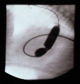
Partially inflated balloon with indentation at the stenosis.
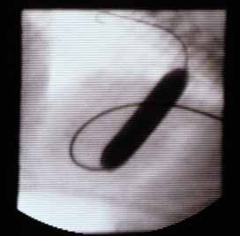
Fully inflated balloon — stenosis opened; indentation gone.
Video of Balloon Valvuloplasty
The pressure gradient was measured immediately after balloon valvuloplasty and showed no change. However, an echocardiogram showed a more open valvular region, and the infundibular muscle appeared to obliterate the right ventricular outflow tract in systole. A continuous wave Doppler taken about 30 minutes after valvuloplasty (below) revealed the gradient was decreasing (55 mmHg) and the waveform had the characteristic shape of a dynamic stenosis — increasing to a higher peak late in systole. This is characteristic of a “suicide” ventricle: when stenosis is relieved, afterload decreases, allowing the ventricle to contract to a smaller diameter, and the tremendous hypertrophy in the right ventricular outflow tract collapses on itself producing a dynamic or functional stenosis in late systole. In some cases this resolves on its own; in others, beta-blocker administration can alleviate the problem by decreasing contractility in this region.

CW Doppler 30 minutes post-valvuloplasty. Pressure gradient decreased to 55 mmHg with a late-peaking waveform characteristic of dynamic (infundibular) obstruction.