Case 7 — Follow-Up
Cindy recovered reasonably well from anesthesia. She had isorhythmic AV dissociation for several hours while recovering but was clinically stable. Echocardiography was repeated the following day when she was fully recovered from anesthesia.
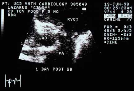
Pulmonic valve after valvuloplasty. Unlike the pre-intervention image, a readily demonstrable orifice is now visible. RVOT — right ventricular outflow tract; PA — pulmonary artery; AO — aorta.
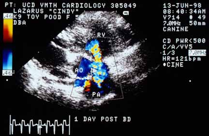
Color flow Doppler after valvuloplasty. The abnormal signal appears primarily due to aliasing rather than turbulence, and is much wider at the pulmonic valve than before intervention. RV; AO; PA.

CW Doppler post-valvuloplasty. Pressure gradient reduced to 43 mmHg (from 210 mmHg). Upward arrow: flow during atrial systole (forceful contraction). Downward arrow: low-velocity pulmonic regurgitation in diastole (~1.6 m/s, consistent with a normal low diastolic pressure gradient).
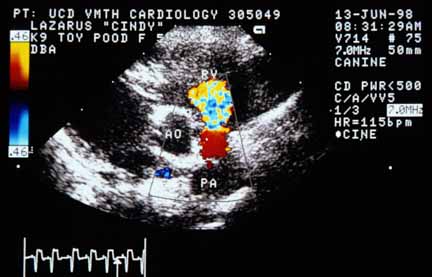
Color flow Doppler demonstrating pulmonic regurgitation in diastole. Laminar flow in the PA (red) aliases at the valve and projects into the RV. The jet appears larger post-procedure but is hemodynamically insignificant due to the low systolic pressure gradient.
In addition to the above studies, another contrast echocardiogram was performed the day after valvuloplasty. It showed almost no bubbles traversing the patent foramen ovale. The dog appeared brighter, and when the owner picked up the dog, it became very excited. Whereas before the intervention the dog became cyanotic and dyspneic, it was able to tolerate the excitement very well. When the owner was called the following day, she was ecstatic — she claimed that the dog was “300% better” than before. Cindy was able to play normally without fatigue, cyanosis, or dyspnea.
The clinical improvement in this dog was almost certainly due to a decrease in hypoxemia brought about by a decrease in right-to-left shunting across the large patent foramen ovale. This decrease probably occurred because the amount of tricuspid regurgitation decreased once the pulmonic stenosis was relieved. Once the resistance to flow through the pulmonic valve decreased, flow would have increased into the pulmonary artery and would have decreased through the fixed resistance in the tricuspid valve. This is much like taking a river with two branches in which one branch has a dam with little flow and blowing up the dam — more flow goes through the branch with the blown-up dam and less through the other. The increase in pulmonary blood flow in this dog probably also improved systemic arterial oxygenation.