Case 11 — Case Discussion
The heart was markedly enlarged on the thoracic radiographs and the left atrium was noticeably larger than all other chambers. On his ECG, he had occasional premature complexes (4th and 25th complexes). These QRS complexes were abnormally oriented, wider than the sinus QRS complexes, and had abnormal T waves opposite in polarity — consistent with premature ventricular complexes (PVCs). Note that each had a P wave in front, but the PR interval was very short compared to normal complexes, leading one to surmise the P waves and QRS complexes were not associated. The premature complexes came in 7 mm (0.28 seconds) after the sinus QRS complexes, depolarizing at a rate of 215 beats/minute.
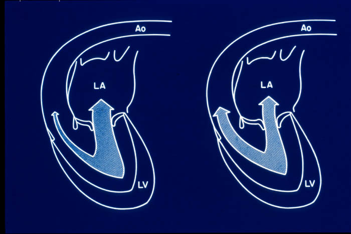
On the echocardiogram, Joey had severe mitral regurgitation due to myxomatous degeneration of his mitral valve leaflets and chordae tendineae. The latter resulted in rupture of at least one minor chord. The ruptured chord was evident on the two-dimensional echocardiograms (the arrow). The force of the jet against the interatrial septum and/or the high left atrial pressure resulted in an endocardial tear and perforation of the interatrial septum — an acquired atrial septal defect. Joey was lucky: a perforation in any other part of his atrium would have resulted in hemorrhage into the pericardial sac and acute cardiac tamponade, most commonly resulting in acute death.

Joey most likely went into right heart failure because of the acquired atrial septal defect coupled with existing tricuspid regurgitation. The increase in right heart volume from the left-to-right shunting at the atrial level would increase right atrial and right ventricular diastolic pressures, resulting in ascites.
The CW Doppler showed a systolic LA pressure of ~55–60 mmHg (calculated two ways: 170 − 115 = 55 mmHg from the MR jet; 47 + 13 = 60 mmHg from the ASD jet). This tremendous increase may explain why the left atrial wall tore and then ruptured.
Treatment
Joey was sent home on furosemide 50 mg 1 tab TID and enalapril 10 mg ½ tab SID.
Discharge Instructions
- Joey has advanced degeneration of the mitral and tricuspid valves causing blood to flow backward in the heart, resulting in pulmonary edema and ascites. He also has an acquired atrial septal defect — a hole in the wall between two heart chambers.
- Lasix 50 mg, one tablet three times daily.
- Enalapril 5 mg (½ of a 10 mg tablet) once daily.
- A blood sample has been taken to check kidney function; results to be called in.
- Recheck next week; he may be started on amlodipine to lower blood pressure and improve heart function.
Follow-Up: One Week Later
Joey was feeling better. He still had some labored breathing but seemed more comfortable. He was eating and drinking well and taking his medications. No coughing, wheezing, or vomiting. Some diarrhea yesterday. No collapsing episodes. Chemistry panel was normal.
Follow-Up: Two Weeks Later
Joey seemed to be losing more weight and was behaving like he was having trouble seeing at night. His appetite was good though he was a picky eater. He had a painful distended abdomen with a fluid wave and an enlarged liver.
Blood pressure: Prior to amlodipine dose, 150/92 with pulse rate 170 bpm. Two hours after amlodipine (0.625 mg), blood pressure decreased to 118/71. The dosage was sufficient to lower BP without hypotension. Joey was maintained on amlodipine ¼ tab (0.625 mg) BID to reduce afterload and thus reduce the regurgitant fraction. Radiographs showed a slight increase in pulmonary infiltrates. Approximately 1 L of modified transudate was tapped from the abdomen.
Conclusion: Joey may need abdominocentesis in the future again. Removal of the ascitic fluid may improve his appetite. Joey will return for a recheck if his respiratory rate increases, if the ascites returns, or if he develops other problems.