Case 17 — Case Discussion
This dog had a left-to-right shunting patent ductus arteriosus (PDA), a congenital and heritable cardiac abnormality. In the dog, it has been proven in miniature poodles that the lesion is caused by a lack of smooth muscle in the ductal wall. Usually at the aortic end there is little to no smooth muscle while at the pulmonary artery end there is enough to result in partial closure, creating a funnel shape. The narrowed end creates resistance to flow and causes high-velocity flow into the pulmonary artery. Since aortic pressure is always greater than pulmonary artery pressure, flow is continuous with peak flow in systole.
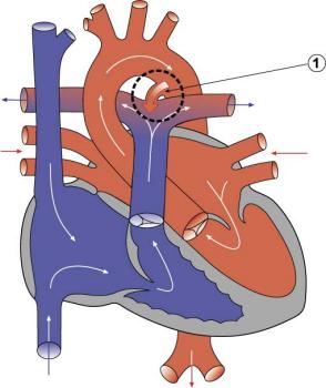
The diagnosis is usually suspected by identifying the characteristic continuous heart murmur, heard best cranially along the left thorax and in the left axillary region. Chico’s murmur had been missed for a year probably because no one had listened this far cranially. Luckily, his PDA was moderate in size so he did not develop fulminant heart failure before diagnosis.
Chico had a classic descending aortic bulge (“ductus bump” or “ductal aneurysm”) on his DV radiograph and a tall R wave in lead II indicating left ventricular enlargement. Echocardiography revealed volume overload of the left ventricle and left atrium and a markedly enlarged main pulmonary artery. The PDA was visualized on 2D and color flow Doppler. The CW Doppler showed a peak velocity of approximately 5 m/s (PG ~100 mmHg), ruling out pulmonary hypertension and concomitant congenital abnormalities.