Case 17 — Intervention (Coil Embolization)

Instead of correcting the PDA surgically, it was closed using two small stainless steel coils covered with synthetic fibers to stimulate thrombosis.

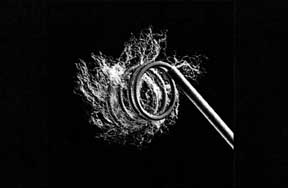
Photograph of the stainless steel coils used for PDA embolization, covered with synthetic fibers.
Coils used for embolization.
Close-up of a coil being deployed from a catheter.
Coil emerging from a catheter.

Chico was anesthetized and transesophageal echocardiography was performed.

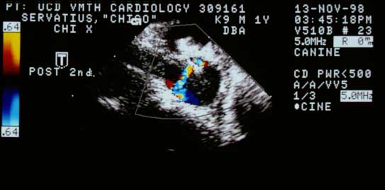
Transesophageal echocardiogram showing the pulmonary artery (PA) end of the PDA (D). The opening was 3.5 mm wide.
Transesophageal echocardiogram. PA end of the PDA (D): 3.5 mm opening.

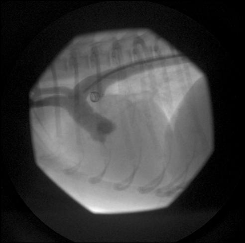
A 5 French catheter was passed via the left femoral artery into the proximal descending aorta. An angiogram was taken to visualize the PDA location.
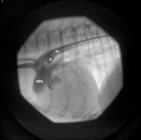
Angiogram showing the PDA location (arrow) between the aorta and main pulmonary artery.
Angiogram showing PDA (arrow).
Labeled angiogram. MPA — main pulmonary artery; D — ductus; Ao — aorta.
Labeled. MPA; D (ductus); Ao.

Video of the angiogram


Two coils were placed: an 8 mm × 5 cm Cook non-detachable coil first, then a 5 mm × 5 cm coil on top.

Fluoroscopic image showing the first coil deployed in the ductus.
First coil deployed in the ductus.
Recording showing ECG on top and systemic arterial blood pressure on the bottom. Diastolic pressure increased immediately after coil placement as the PDA was occluded.
Diastolic pressure increased immediately after coil placement as the PDA was occluded.
Post-coil transesophageal echocardiogram showing the coil and a large thrombus (arrow) in the pulmonary artery end of the ductus arteriosus.
Post-coil TEE. Coil and large thrombus (arrow) in the PA end of the ductus.
Post-coil color flow Doppler showing two small residual jets through the thrombus and ductus.
Post-coil color flow Doppler. Two small residual jets through the thrombus.
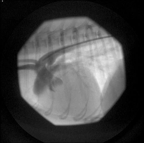
Post-procedure angiogram showing coils in the ductus with no contrast in the pulmonary artery.
Post-procedure angiogram. Coils in the ductus; no contrast in the PA.

Video of angiogram following coil deployment
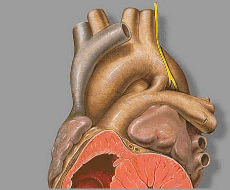
Diagram illustrating coil embolization of a PDA.
Diagram of a PDA.