Case 12 — Two-Dimensional Echocardiograms

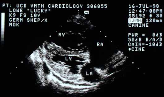
Right parasternal long-axis view showing remarkable RV and RA chamber enlargement. The LV and LA chambers are smaller than normal. The RV free wall is thicker than normal.
Right parasternal long-axis view. Remarkable RV and RA enlargement. The LV and LA are smaller than normal. The RV free wall is thickened.
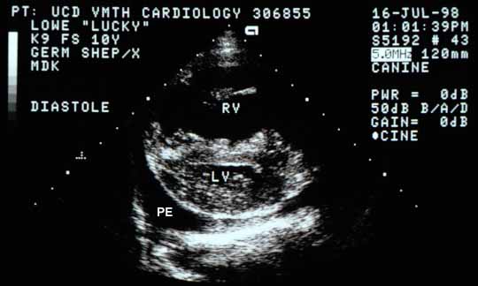
Cross-sectional view of the ventricles in diastole. The RV chamber is very large and the LV chamber is small. The interventricular septum is flattened, suggesting diastolic RV pressure is higher than LV pressure. PE — pleural effusion.
Cross-sectional view (diastole). RV is very large; LV is small. IVS is flattened, suggesting RV diastolic pressure exceeds LV diastolic pressure. PE — pleural effusion.
Same cross-sectional view in systole showing the LV becomes round again as septal flattening disappears. The LV chamber is ~18 mm wide (normal ~35 mm). Chamber volume is approximately 1/7 of normal.
Same view (early systole). LV becomes round as septal flattening resolves. LV width ~18 mm (normal ~35 mm); estimated volume ~1/7 of normal.
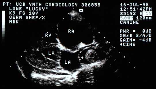
Longitudinal section showing the four cardiac chambers and their relative sizes. A large mass (arrow) is visible in the right caudal lobar pulmonary artery branch, which is also markedly enlarged.
Longitudinal section. A large mass (arrow) is visible in the markedly enlarged right caudal lobar pulmonary artery branch.
Quiz

1. The large right ventricular chamber and thickened right ventricular free wall are characteristic of what hemodynamic abnormality?

2. The higher than normal diastolic right ventricular pressure suggested in the second image will cause what?

3. What do you think the mass in the pulmonary artery is in the last image?