Case 12 — Electrocardiogram

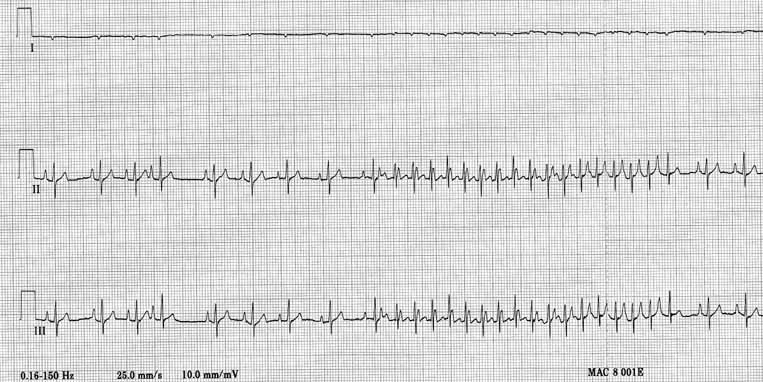
Three-lead ECG (I, II, III) showing deep S waves in leads II and III. Lead I QRS is completely negative though very small. All complexes are small, likely due to pleural effusion. Basic rhythm is sinus at ~100-120 bpm. The fourth complex is abnormal. After the ninth complex, a period of nonsustained tachycardia occurs.
Three-lead ECG (I, II, III). Small complexes (pleural effusion). Deep S waves in II and III with negative QRS in lead I indicate RV enlargement. Sinus rhythm ~100–120 bpm with a bout of nonsustained tachycardia after the 9th complex.

Quiz

1. There is evidence of one chamber being enlarged. Which one?

2. The complexes are all small. Which of the following is not a possible cause for this in this dog?

3. This dog has a bout of tachycardia. What type of tachycardia is it?