Case 12 — Echocardiograms of the Pulmonary Arteries

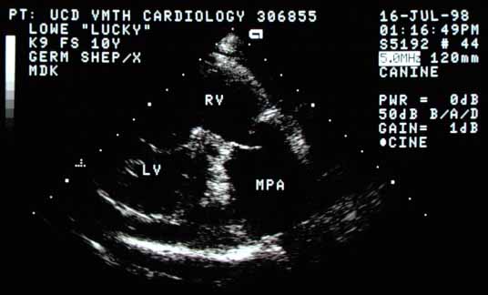
Non-standard echocardiographic view of the right ventricle (RV) and main pulmonary artery (MPA). The MPA is markedly enlarged, suggesting very high pressure or flow. One pulmonic valve cusp is open and appears moderately thick.
Non-standard view of the RV and main pulmonary artery (MPA). The MPA is markedly enlarged. A pulmonic valve cusp appears moderately thick.
Right parasternal cross-sectional view of the base of the heart. The main pulmonary artery diameter is much larger than the aortic root (Ao). The pulmonic valve cusps look moderately thick. The right caudal lobar pulmonary artery (RPA) is markedly enlarged. The same mass seen previously (arrow) is at the terminal portion of the RPA. RVOT — right ventricular outflow tract.
Cross-sectional base view. The MPA diameter far exceeds the Ao. The RPA is markedly enlarged with the same mass (arrow) at its terminal portion. The mass was not mobile. RVOT.