Case 12 — Thoracic Radiographs

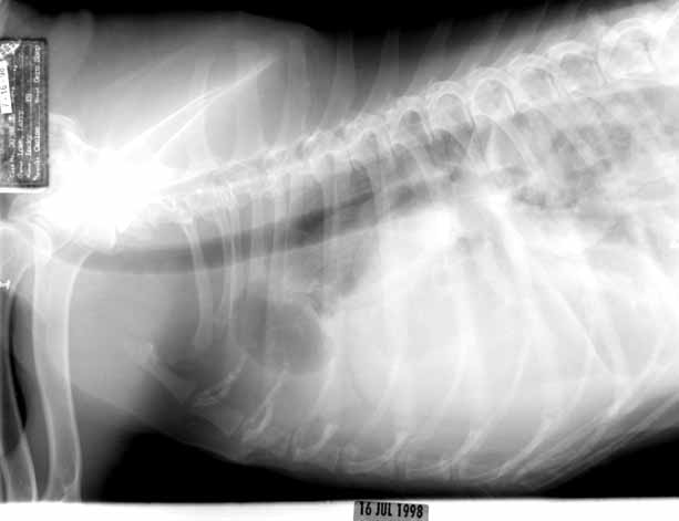
Lateral

Lateral thoracic radiograph showing an obscured cardiac silhouette due to pleural effusion. Pulmonary arteries are visible.
Lateral thoracic radiograph.

DV

DV thoracic radiograph showing an obscured cardiac silhouette due to pleural effusion.
DV thoracic radiograph.

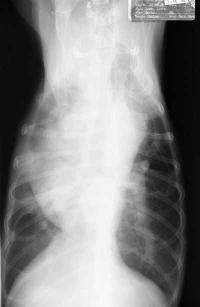
VD

VD thoracic radiograph showing the cardiac silhouette more clearly than the DV or lateral views. The main and left caudal lobar pulmonary arteries are large. The left caudal artery is also tortuous.
VD thoracic radiograph. The cardiac silhouette is more visible in this view.

Quiz

1. Why is the cardiac silhouette obscured on the lateral and DV views but not the VD view?

2. How would you characterize the pulmonary vasculature?