Case 12 — Case Discussion
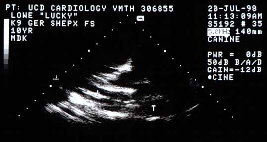
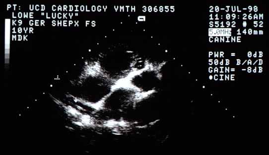
This dog has pulmonary thrombosis or thromboembolism based on the large mass compatible with a thrombus on the echocardiogram. Although this mass could theoretically be something other than a thrombus, it is extremely unlikely. The mass is round, smooth, and homogeneous — all characteristics compatible with a thrombus.
There are numerous reasons for a dog to have pulmonary thromboembolism, including hyperadrenocorticism, diabetes mellitus, autoimmune hemolytic anemia, and protein-losing glomerulonephropathy. Basic blood work ruled out everything except the last. A urine protein-to-creatinine ratio was 5 (normal <1), confirming protein-losing glomerulonephropathy. However, plasma antithrombin III concentration was 88% of control (normal >80%), ruling this out as the cause.
Because common causes of pulmonary thromboembolism had been ruled out and the radiographic appearance of the right caudal lobar pulmonary artery was very suggestive of heartworm disease, Lucky was treated for adult heartworms with melarsomine. At a follow-up echocardiogram 4 days later, she had more pleural effusion and the right caudal lobar pulmonary artery distal to the thrombus could be visualized.
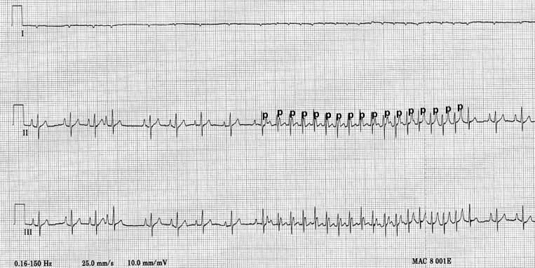
Lucky also had a supraventricular tachycardia (SVT) that was somewhat irregular, with identifiable P waves before each QRS complex. The atrial rate was not regular, and the PR interval was also irregular, suggesting the SVT may have been due to an automatic (rather than reentrant) focus, most likely in the right atrium given the RA enlargement.
Follow-Up
Because several heartworm antigen tests were negative, Lucky most likely only had a couple of male worms. The heartworm antigen tests available only detect an antigen from the female reproductive tract and eggs, so male worms go undetected. Lucky clearly had one worm in her right caudal pulmonary artery branch and radiographic evidence of another in the left caudal lobar artery branch (enlarged and tortuous on radiographs).
Because she already had severe pulmonary and cardiac disease (heartworm disease class 3), Lucky was treated conservatively with one injection of melarsomine. One injection primarily kills male worms. The owner was instructed to return in 4 weeks for two injections 24 hours apart. Lucky was sent home on increased Lasix (25 mg q8h) and enalapril (10 mg q12h). Ten days later she required another 1500 ml thoracocentesis and the Lasix was increased to 37.5 mg q8h. Digoxin was discontinued due to suspected toxicity (anorexia that resolved within 48 hours), then restarted at 0.0625 mg q24h. Diltiazem was started at 15 mg q8h for the SVT, eventually increased to 30 mg (~2 mg/kg) q8h, which regularized her rhythm to ~160 bpm.
Three weeks after melarsomine, Lucky was lethargic with poor appetite and rear-leg weakness. She had 1.3 liters of pleural fluid drained. The heartworm was no longer visible on echocardiogram but the thrombus had enlarged rather than decreased in size. Her hematocrit had fallen from 44 to 30, BUN increased from 40 to 75, ALT from 52 to 163, and WBC from 25,000 to 39,000.
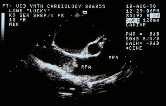
Options including thrombolytic therapy and surgical removal of the thrombus were discussed with the owner. He decided to treat Lucky conservatively at home in the hope that her fibrinolytic system would break down the clot. The owner phoned one week later to let us know that Lucky died in his arms. He buried Lucky in his yard.